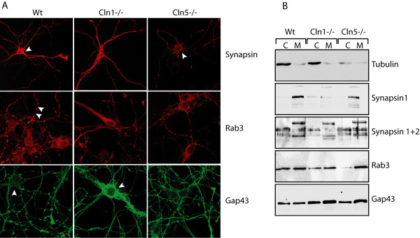
Figure 5.
A) Immunofluorescence analysis of Synapsin, Rab3 and GAP-43 staining in E15 primary cortical neurons of wt, Cln1-/- and Cln5-/- mice.B) Western blot analysis of cytoplasmic and membrane bound fractions of cytoskeletal, growth-cone and synapse assembly proteins. Antibodies: β-tubulin, synapsin 1, synapsin 1&2, Rab3 and Gap43.

