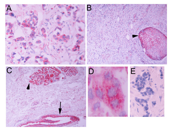
Figure 2.
APRIL immunoreactivity in invasive breast carcinoma (A), DCIS component of the same carcinoma (B, arrowhead), and normal looking ducts (C, arrow) and lobules (C, arrowhead). In D, a higher magnification is shown. APRIL immunoreactivity is presented as discrete cytoplasmic dots. E: Normal Ig isotype staining.

