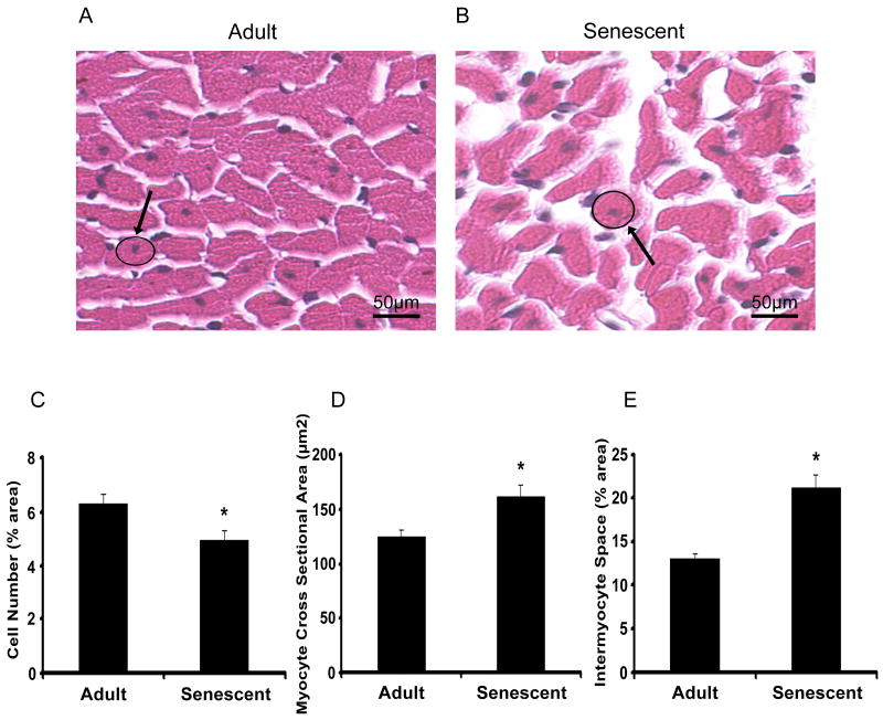
Figure 2.
H&E staining images in paraffin sections of adult (n=15) and senescent mice (n=14) were scanned at a magnifications of 40x and 100x. Myocyte cross sectional areas were calculated from circumstance of myocyte with round and central nuclear measured with the software of Echo-Station. Compared with adult group, myocyte cross sectional areas increased 40% in senescent group (p<0.05) (A). Quantitation of the percentage of white space to the total stained area and blue color staining (hematoxylin) to the pink (Eosin) were performed. Intermyocyte space increased 62% in senescent group compared with adult group (p<0.05) (B). Myocyte cell numbers decreased in senescent mice (C).

