
Fig. 1. Age-dependent loss of repressive histone methylation in oligodendrocyte lineage cells.
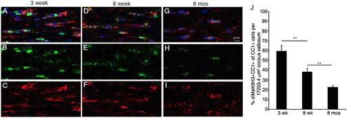
High magnification view of representative confocal imaging of the medial corpus callosum in coronal brain sections from 3-week (panel A-C), 8-week old (panel D-F), and 8-month (panel G-I) old mice. Sections were immunostained with antibodies against oligodendrocyte surface marker CC1 (red in panel A, C, D, F, G, I) and against dimethylated lysine 9 residues on histone H3 (green in panel A, B, D, E, G, H). Nuclei were visualized with DAPI (blue). Note the progressive decline of immunoreactive nuclei for repressive histone methylation (dimethylated lysine 9 residues on histone H3) occurring as part of the aging process. The bar graphs (panel J) indicate the percentage of Methyl-K9-H3+/CC1+ cells normalized by the total number of CC1+ cells (** p<0.01). (Scale bar= 10 μm, 63X objective).