
Figure 1.
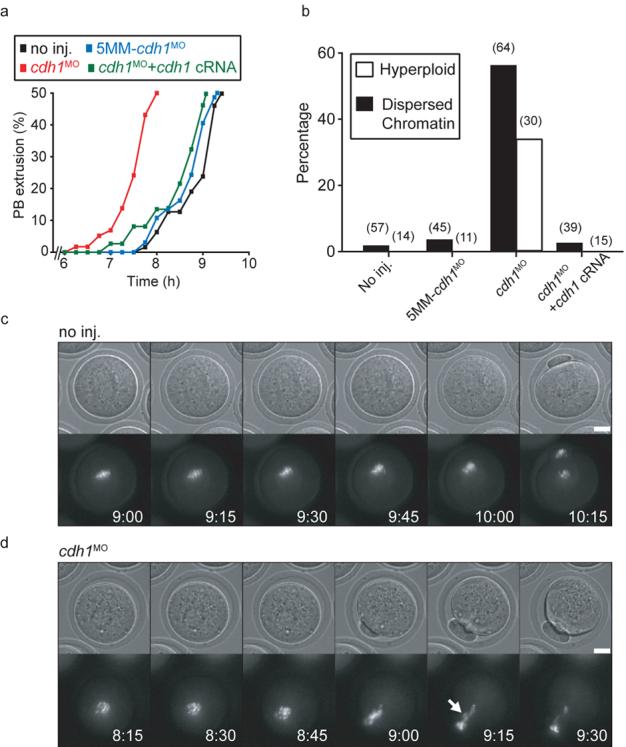
Cdh1 loss accelerated meiosis I leading to premature homologue segregation and non-disjunction. a, Timecourse of PB extrusion rates in oocytes injected with cdh1MO (n=58), 5MM-cdh1MO (n=37), cdh1MO and cdh1 cRNA (n=67); or no injections (n=64). Cdh1 knockdown accelerated PB extrusion. b, Percentages of dispersed chromatin and hyperploidy observed in matured non-injected control oocytes, or those injected with 5MM-cdh1MO, cdh1MO and cdh1 cRNA as stated. Number of oocytes examined in parentheses. c and d, Chromatin (Hoechst) and brightfield images of maturing non-injected oocytes (c) and those injected with cdh1MO (d). Cdh1 knockdown oocytes had no discernible congression of chromosomes on a metaphase I plate and had lagging chromosomes during anaphase (arrow). (c) and (d) time stamp is h:min, scale bar is 20μm. (c) and (d) are selected frames from Supplementary Movies 1 and 2. (a), (c) and (d) times are relative to GVB.