
Figure 2.
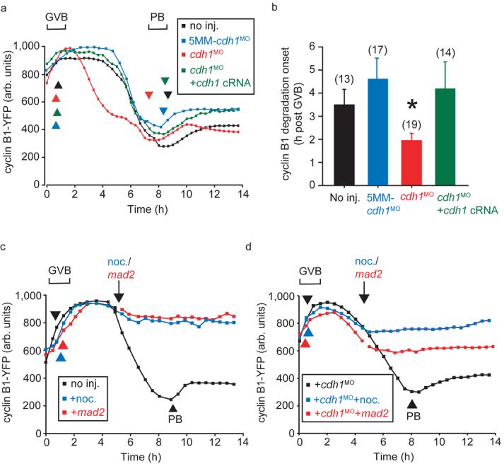
Premature cyclin B1 degradation following cdh1 knockdown. a, Cyclin B1-YFP fluorescence during maturation of oocytes injected with cdh1MO, 5MM-cdh1MO and cdh1 cRNA as stated. b, Calculated from (a), the timing of initiation of cyclin B1-YFP degradation after GVB (asterisk indicates P=0.034, significantly different from non-injected, ANOVA). The error bars represent s.d. Numbers of oocytes in parentheses, pooled for each condition from at least 2 independent experiments. c, Cyclin B1-YFP fluorescence in oocytes during maturation, at the arrow when indicated oocytes were injected with mad2 cRNA or nocodazole was added to the medium. d, As for (c), in oocytes following cdh1 knockdown. Mad2 and nocodazole both inhibited PB extrusion, which has been marked in control oocytes by the arrowheads. (c) and (d) are representative recordings of at least 12 oocytes.