
Figure 3.
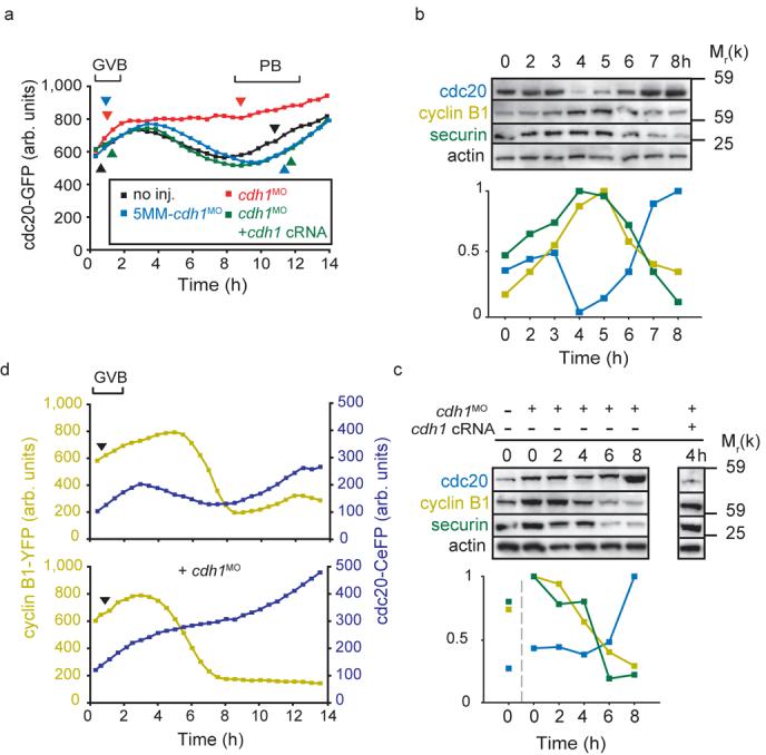
Cdc20 degradation precedes that of cyclin B1 and securin during meiosis I. a, Representative cdc20-GFP fluorescence recordings in oocytes microinjected with cdh1MO (n=16); 5MM-cdh1MO (n=23), cdh1MO and cdh1 cRNA (n=14); or no further injections (n=22). Note that cdh1 knockdown prevented cdc20-GFP degradation between GVB and PB. (b and c), Western blot and densitometric analysis of endogenous cdc20, cyclin B1 and securin levels (50 per lane) at the times indicated relative to GVB (0 h is GV arrest). In (b) oocytes were uninjected; note the early loss in cdc20 at 4h, ahead of both cyclin B1 or securin. In (c) oocytes were microinjected with cdh1MO and cdh1 cRNA as indicated; note that cdh1 knockdown abolished cdc20 loss and brought forward cyclin B1 and securin degradation. Cdc20 degradation at 4 h was recovered by cdh1 cRNA rescue. Actin was used as a loading control. Blots are representative of 2 further replicates. d, Cyclin B1-YFP and cdc20-CeFP fluorescence levels recorded simultaneously during maturation, with or without microinjection of cdh1MO as indicated. Representative traces (top, n=40; bottom n=44) of cyclin B1-YFP and cdc20-CeFP fluorescence during maturation. In (a) and (d) the timing of GVB and PB extrusion are marked by arrowheads. (b) and (c) Uncropped Westerns for cdc20, securin and cyclin B1 are displayed in the Supplementary Information, Fig S4.