Figure 4.
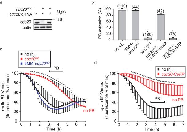
Essential role of cdc20 in meiosis I. a, Western blot of matured oocytes (50 per lane) following injection of cdc20MO or cdc20 cRNA as indicated at the GV stage. The morpholino prevented re-accumulation of cdc20 during MI. The uncropped Western is displayed in the Supplementary Information, Fig S4. b, PB extrusion rates following maturation in oocytes injected with cdc20MO, 5MM-cdc20MO, cdc20 cRNA, or cdc20-GFP cRNA as stated. Cdc20 knockdown blocked completion of MI which was rescued by cdc20 cRNA. c, Cyclin B1-Venus degradation rate, plotted with respect to time as a percentage of the maximum recorded fluorescence, in oocytes injected with cdc20MO (n=18); 5MM-cdc20MO (n=15); or without further microinjection (n=15), pooled for each condition from at least 2 independent experiments. d, Cyclin B1-Venus degradation rate plotted as for (c), in cdh1 knockdown oocytes injected cdc20-CeFP cRNA (n=38) or no further injections (n=34), pooled for each condition from at least 2 independent experiments. The pipette concentration of cdc20-CeFP was high, four-fold that used in any other experiment. In (b), (c) and (d), asterisks imply significantly different from non-injected group, (p<0.05; b, ANOVA; c and d, t-test). In (c) and (d) the range over which PB extrusion occurred is as marked.

