
Fig. 1. FoxI1e is expressed in a dorsal to ventral wave in the blastula and gastrula stages.
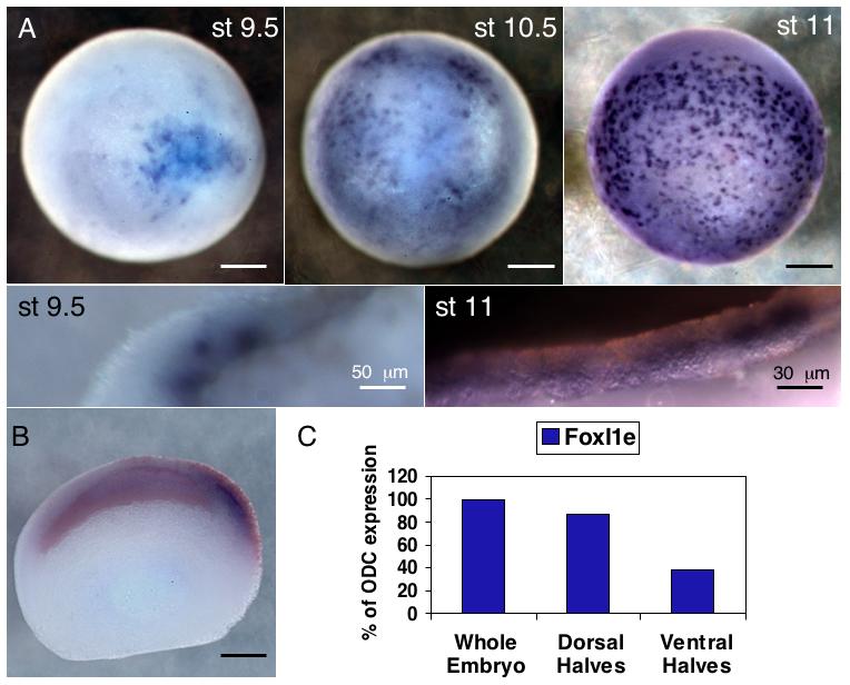
(A) In situ hybridization for FoxI1e shows initial staining at stage 9.5 on one side of the embryo, and then spreading across the embryo. Its expression is always mosaic. (B) Embryos injected in the two dorsal, animal blastomeres at the 4-cell stage with 50 pg of β-Gal mRNA were stained with Red-Gal before in situ hybridization, showing the initial expression is on the dorsal side of the embryo. (C) Embryos were dissected into dorsal and ventral halves at stage 10 and frozen for real-time PCR. FoxI1e expression is enriched on the dorsal side at stage 10. Results are normalized to ODC expression levels. (D) Stage 11 embryos were stained for FoxI1e and sectioned. Staining with Wheat Germ Agglutinin defines a small population of FoxI1e positive cells between the sensorial and epithelial layers of the ectoderm. Scale bars represent 200 μm, unless otherwise noted.