
Figure 1.
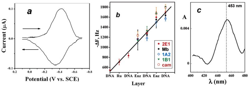
Characterization data for films: (a) Background-subtracted, smoothed reversible CV of cyt P450 1B1 on PG electrode at 50 mV/s in pH 7.1 phosphate buffer + 50 mM NaCl. Film composition on electrode was PDDA/DNA/cyt P450 1B1/DNA. (b) Influence of film layers added on QCM frequency decrease for enzymes used in this study. The first three film layers are omitted (RuPVP/DNA/RuPVP) for clarity. (c) Visible difference spectrum for immobilized PDDA/cyt P450cam film on glass microscope slide after reduction to the ferrous form and addition of CO showing the characteristic cyt P450 FeII-CO band near 450 nm.