
Figure 3.
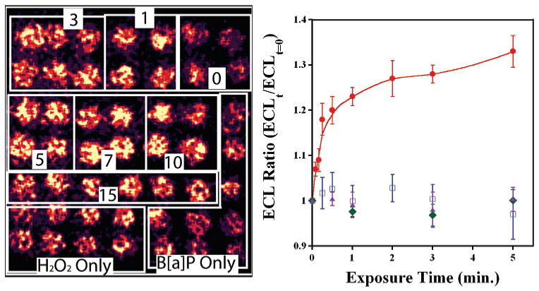
(a) CCD image of ECL array with 49 individual RuPVP/DNA/enzyme spots containing cyt P450 1B1. Boxes denote spots that were exposed to 0.5 mM H2O2 + 100 μM B[a]P for the labeled times (min). Controls on bottom were exposed to B[a]P or H2O2 only for increasing amounts of time from 1 to 7 min as viewed from right to left (not marked for clarity). (b) ECL ratio plot demonstrating the increase in ECL intensity vs time of enzyme reaction. Controls show ECL response vs time exposed to B[a]P only (blue squares), H2O2 only (green diamonds), and 0.5 mM H2O2 + 100 μM B[a]P + 30 μM of inhibitor αNF (purple triangles).