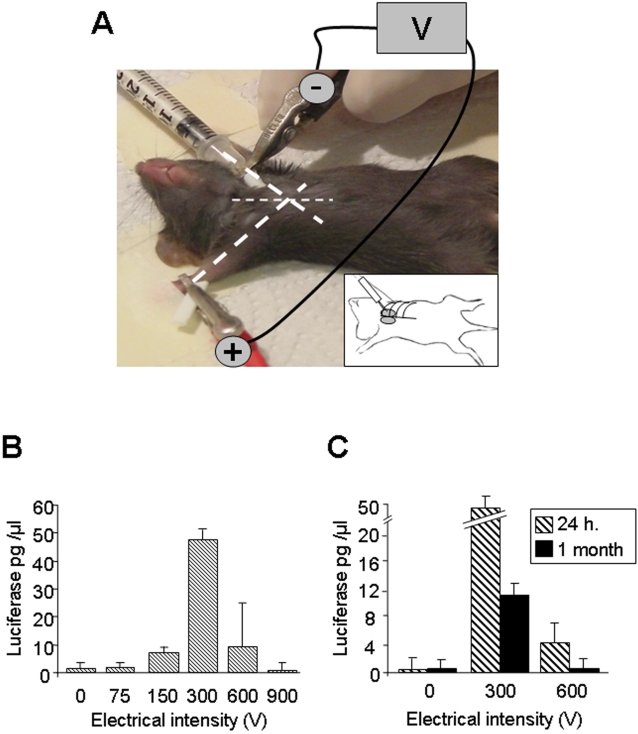
Figure 1. Monopolar electroporation of the thymus in anaesthetized mice.
(A) Bold dotted lines indicate the angle of axis legs. The needle (optimal size: 3 mm) is inserted between the two first ribs, under the ribcage, following the angle of the paw parallel to the benchtop. The stimulating syringe, delivering electric pulses and plasmid DNA, represents the anodic electrode. The cathodic pole is represented by a crocodile clip attached to the paw of the side opposite to the injection point. The inset represents a schematic representation of the needle insertion through the ribcage to inject the thymus. (B) Luciferase activity in the thymus 24 hours after injection with or without electroporation. 10 µg of luciferase DNA plasmid in a volume of 10 µl was injected into each thymic lobe of 10 mice for 0 V, 10 mice for 300 V and 5 mice for all other voltages. Whatever the electric intensity applied, all mice survived. (C) Luciferase activity 24 hours (h) and one month after injection in the thymus without or with electroporation at 300 and 600 V, respectively. Four mice were analyzed for each condition.

