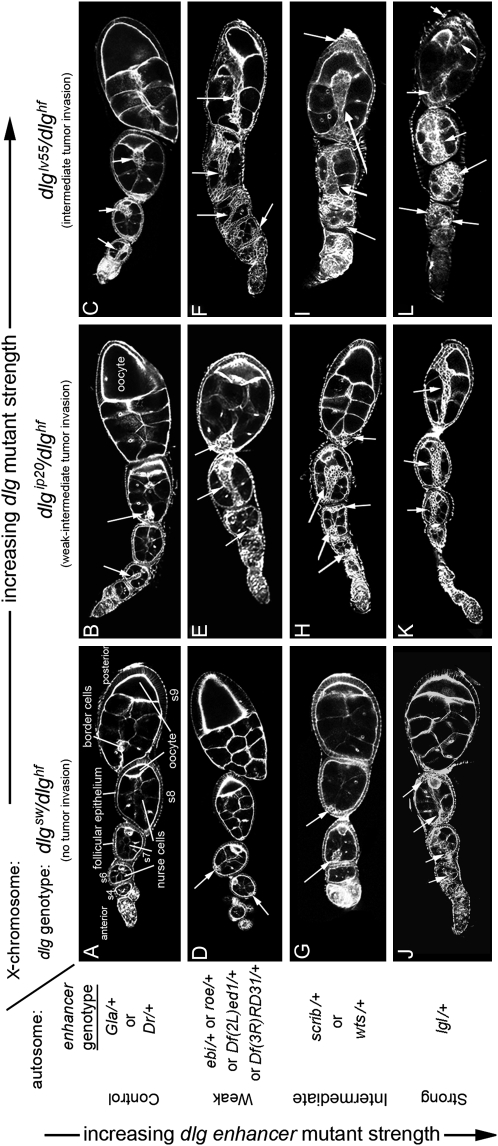
Figure 2.—
Characterization of dlg enhancer phenotypes. A–L are organized horizontally left to right by increasing dlg strength (X chromosome, dlg genotype), and vertically top to bottom by increasing dlg enhancer strength (autosome, enhancer genotype). Strings of maturing egg chambers that are representative of the indicated X chromosome; autosome genotype are shown (arrows point to tumors). (A) Control egg chambers of genotype dlgsw/dlghf; Gla/+ appear wild type. Stage 4 (s4) to s9 egg chambers are evident. Each egg chamber has 16 germ cells, 15 anterior nurse cells, and a posterior oocyte. A follicular epithelium surrounds each egg chamber. A small cluster of cells called border cells exit the anterior epithelium at stage 9 and migrates between the nurse cells to the oocyte. (B) Control egg chambers of genotype dlgip20/dlghf; Dr/+. They have small tumors that invade the germ line starting as early as stage 2 of oogenesis (arrows). (C) Control egg chambers of genotype dlglv55/dlghf; Dr/+. They have intermediate-size tumors that invade more frequently than in dlgip20/dlghf; Dr/+ animals (see text). (D, G, and J) All dlg enhancers cause tumors in dlgsw/dlghf animals that typically do not have tumors (compare to control, A). (E, H, and K) All dlg enhancers increase the frequency and size of tumors in dlgip20/dlghf animals. Whereas tumors are observed in ∼66% of dlgip20/dlghf ovarioles, they are observed in 100% of dlgip20/dlghf; enhancer/+ ovarioles, even for the weakest enhancers. (F, I, and L) In dlglv55/dlghf; enhancer/+ animals, the tumors become increasingly larger and more aggressive in appearance with increasing dlg enhancer strength, often appearing to consume the entire egg chamber in the most severe cases (L).

