
Figure 2.
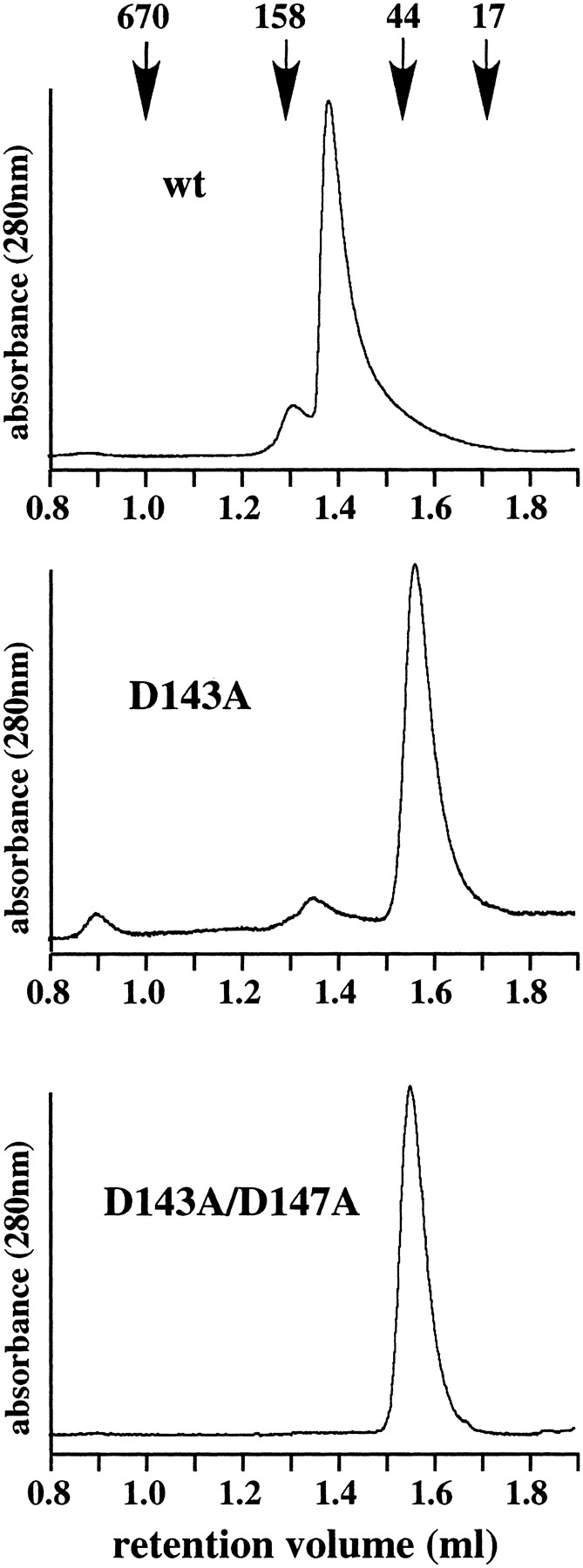
Investigation of trimer formation by gel filtration analysis. The wild-type and mutant PfuPCNA proteins were subjected to a gel filtration column. Elution of the proteins was monitored by absorbance at 280 nm. The molecular masses on the top were from a gel filtration standard (BioRad), containing thyroglobulin (670 kD), γ-globulin (158 kD), ovalbumin (44 kD), myoglobulin (17 kD).