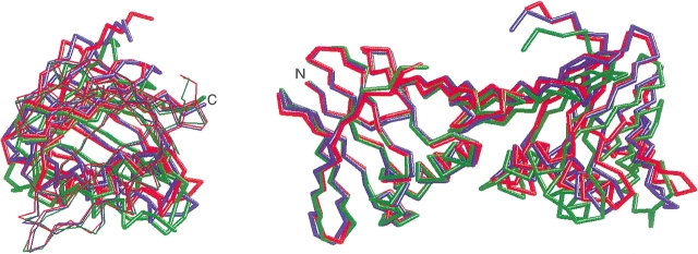
Figure 6.
Domain movement in the mutant PfuPCNA crystals. The monomer structures of the two mutant PfuPCNA crystals were superimposed on the wild-type structure. The wild-type, D143A, and D143A/D147A are indicated in blue, green, and red, respectively. A side view (left) and a top view (right) are shown. In the side view, the N-terminal and the C-terminal domains are shown with a thin and a thick line, respectively.

