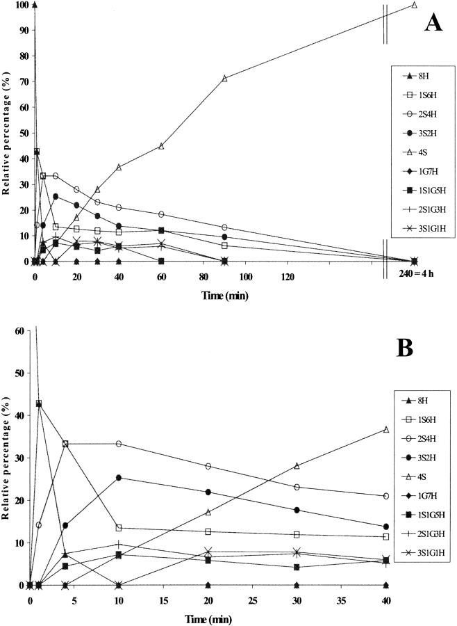
Figure 4.
(A) Time-course analysis of folding in the presence of protein disulfide isomerase (PDI) Δ455–457, the early stages being shown in (B). PDI Δ455–457 was incubated with GSH/GSSG and the intermediates were analyzed as described for wild-type PDI in Figure 3 ▶. For the sake of clarity, error bars are not shown. nS represents intramolecular disulfide bonds, nG mixed disulfides with glutathione, and nH free thiols.

