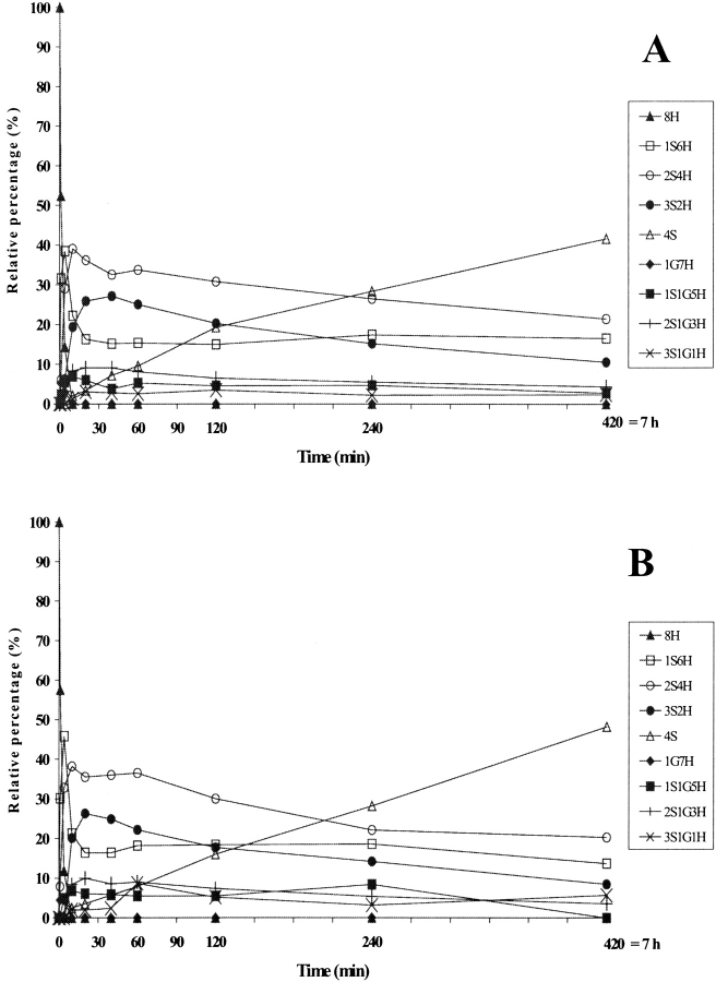
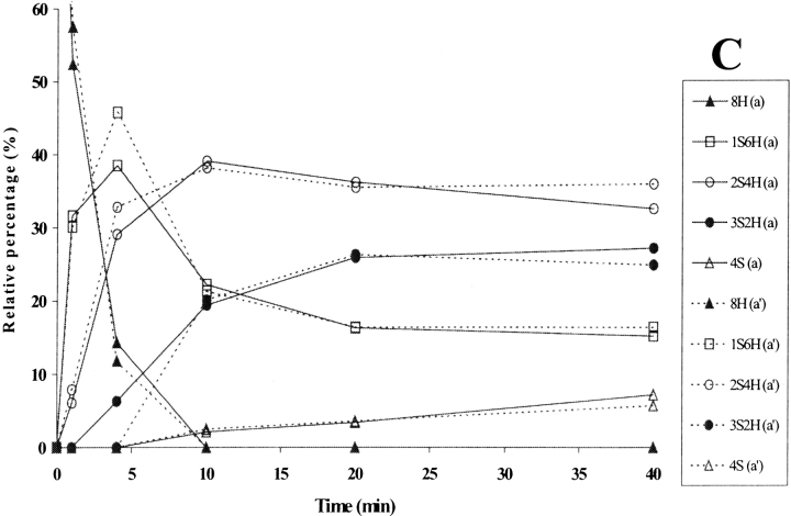
Figure 8.
Time-course analysis of folding in the presence of protein disulfide isomerase (PDI) domain a (A) and a′ (B), the major species accumulating at the early stages being shown in (C). The continuous and dotted lines refer to intermediates present in (A) and (B), respectively. The PDI domains were incubated with GSH/GSSG and the intermediates analyzed as described in Figure 3 ▶. For the sake of clarity, error bars are not shown. nS represents intramolecular disulfide bonds, nG mixed disulfides with glutathione, and nH free thiols.


