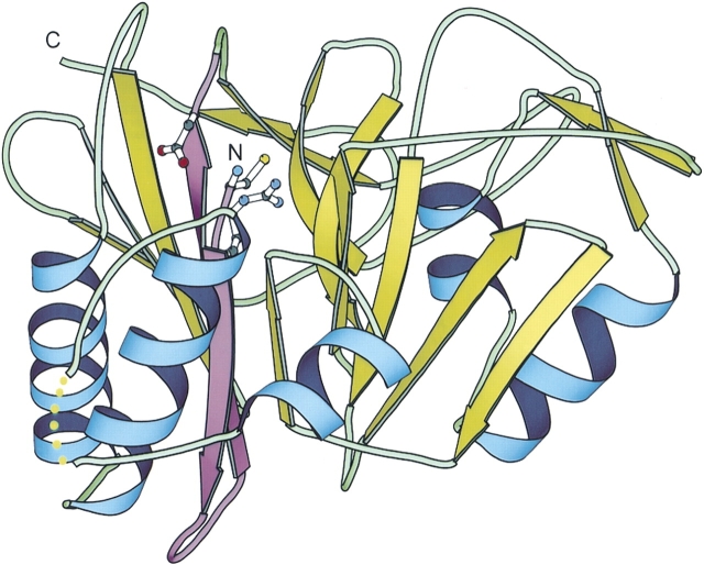
Figure 1.
The structure of the penicillin V acylase from Bacillus sphaericus. The Protein Data Bank (PDB) code is 3pva, chain A. The first β hairpin is highlighted in purple. The backbone amino group and the side chain of Cys 1 and the side chains of Arg 17 and Asp 20 are shown in ball and stick. The structure is drawn using the program BOBSCRIPT (Esnouf 1997).

