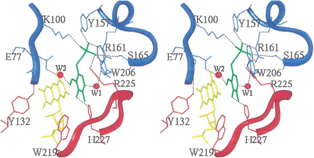
Figure 4.
Stereoview of the active site of human PNPOx, showing the bound PLP ligand (green) at the re face of the FMN cofactor (yellow). The protein residues are colored cyan and red for monomer A and monomer B, respectively. Atoms are shown in stick representation. Salt-bridge and hydrogen-bonding interactions between the PLP and protein are represented by dotted black lines. Water molecules are labeled W1 and W2, and are represented by red spheres. The figure was generated with INSIGHTII (INSIGHT/DISCOVER: Molecular Simulations, Inc.) and labeled with SHOWCASE.

