
Figure 3.
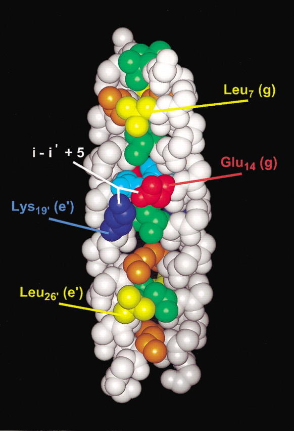
Space-filling representation of the GCN4/cortexillin Hybrid 2 structure. Residues in positions a and d along the hydrophobic core are displayed in orange and green, respectively, with the exception of Asn 15 (a position) in light blue. Leu 7 (g) and Leu 26 (e) side chains are displayed in yellow. Glu 14 (g) and Lys 19′ (e‘) side chains are displayed in red and dark blue, with the i to i‘ + 5 interchain interaction shown in white.