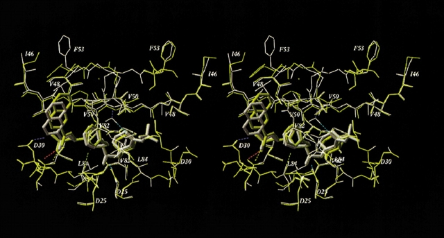
Figure 4.
Superimposed stereo views of the model SQV complexes with the WT (yellow) and the quadruple mutant (M46I/G48V/I50V/I84L, white) HIV-1 PRs. The SQV molecules are shown as tube models. Hydrogen bonds of the ligand and flap water molecule in the mutant complex are displayed. Mutated residues and other important residues are labeled.

