
Figure 4.
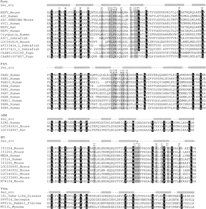
Multiple sequence alignments of the PAAD family. Black background denotes conserved external residues, gray background denotes functional important residues. The "Sec_str" line shows the secondary structure of the reference sequences in each subfamily, namely MEFV_Mouse, PAN2_Human, AIM2_Human, IFI204_Mouse and 18L_Yaba-Like_Disease. The secondary structure was identified from the homology model. The Accession numbers for sequences are the same as Figure 1 ▶.