
Figure 4.
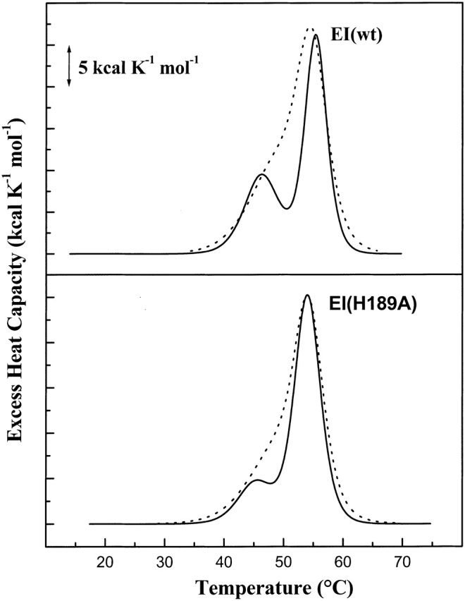
DSC scans of 0.23 mg/mL dephospho-EI(wt) (upper panel) and 0.20 mg/mL EI(H189A) (lower panel) in either Buffer A containing 20 mM phosphate (thick lines) or Buffer B containing 20 mM Hepes (dashed lines) at 30°C/h scan rate and pH 7.5. DSC data are corrected as in Figure 2 ▶.