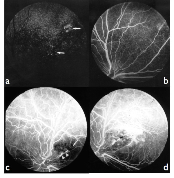
Figure 3.
Fluorescein angiograms: 3a: fundus of the figure 1a, choroidal phase: combined window effect and masking effect (arrows). 3b: fundus of the figure 1a, late venous phase: epithelial fluorescein leakage at the level of lesions. 3c: fundus of the figure 1d, laminar venous phase: masking effect by retinal pigment at the level of the lesion (arrow); linear leakage of fluorescein in the pigment epithelium (points of arrows). 3d: fundus of the figure 1d, late venous phase: most important diffusion of fluorescein at the periphery of the lesion; the hyperfluorescent lines (linear leakage) are corresponding to localized ruptures of the Bruch's membrane.

