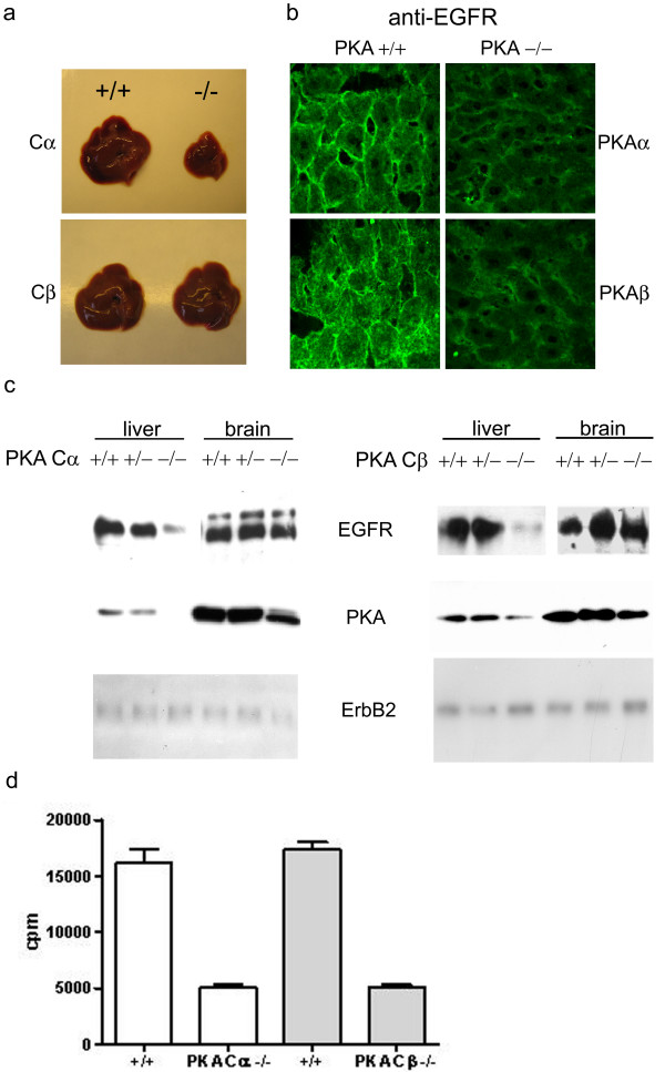
Figure 1.
Effect of PKA Cα or Cβ ablation on EGFR expression. (a) Comparison of liver from wild type and PKA Cα and Cβ ablated mice. PKA Cα KO mice showed a clear uniform reduction in size. (b) Confocal immunofluorescence microscopy of frozen liver sections stained by sheep anti-EGFR and Cy3-conjugated donkey anti-sheep antibodies. (c) Western immunoblotting analysis of EGFR expression in liver and brain from wt (+/+), heterozygote (-/+), and Cα and Cβ KO (-/-) mice. Immunoblots were incubated with sheep anti-EGFR, rabbit anti-pan PKA C and mouse anti-PKA C, and rabbit anti-erbB2. Secondary HRP-conjugated anti-IgG antibodies were used for detection. (d) PKA kinase activity in mouse liver. Activity was assayed by phosphorylation of the PKA-specific substrate Kemptide using γ-[32P]ATP. The assay was performed in the presence of cAMP. Activity was measured by liquid scintillation in 3 ml Opti-fluor. Values are given as counts per minute (cpm).

