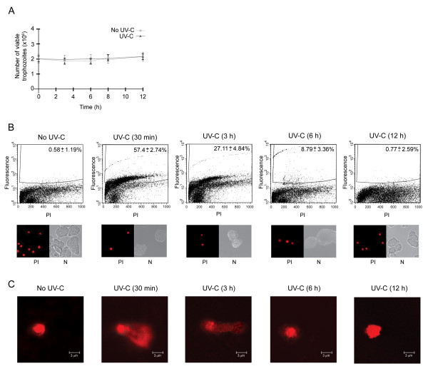
Figure 1.
Effect of UV-C irradiation on cell viability and DNA integrity of trophozoites. A. Growth curves of non-irradiated and irradiated trophozoites (150 J/m2 of UV-C light for 8 s). B. TUNEL assay and flow cytometry (FACS) assays of non-irradiated (No UV-C) and irradiated (UV-C) trophozoites harvested at different times (30 min, 3, 6 and 12 h). Upper panels, histograms show the DNA fragmentation percentage in fluorescence positive cells. The abscissa indicates fluorescence of propidium iodide (PI), and the ordinate indicates fluorescence of Alexa 488-labeled 3' ends of DNA. The number inside each histogram denotes the percentage of fluorescence positive cells above the cut-off line. Lower panels, PI-staining cells were checked in the epifluorescence microscope to confirming the absence of cytoplasmic stain. PI, propidum iodide, N, Nomanski optics. C. Neutral comet assays of non-irradiated (No UV-C) and irradiated (UV-C) trophozoites harvested at different times (30 min, 3, 6 and 12 h). Electrophoretic migration of DNA was from left (anode) to right (cathode).

