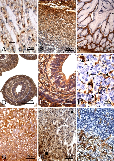
Figure 2.
Fascin distribution in human embryonic, fetal, and normal adult tissue. (A) Vascular endothelial cells (arrows) of the heart showed expression of fascin, whereas cardiocytes were negative at 22 weeks of gestation. (B) Fascin protein was expressed in the elastic membrane of the aorta at 22 weeks of gestation. (C) Glandular epithelium of the intestinal tract was fascin negative at 20 weeks of gestation. (D,E) Immunoreactivity for fascin was shown in cells of the gastrointestinal tract at 8 weeks of gestation. (F) In the liver, expression of fascin was observed in sinusoidal, endothelial (arrows), and Kupffer cells (triangles), whereas normal hepatocytes (arrowheads) were negative at 22 weeks of gestation. (G) Adrenal cortex and medulla showed strong reactivity at 12 weeks of gestation. (H) In the normal adult adrenal gland, fascin immunoreactivity was exhibited in cells of the zona glomerulosa (rectangle) and decreased in the zona fasciculate (triangle). (I) Fascin protein was present in follicular dendritic cells of the thymus and absent in the Hassal corpuscle (arrows).

