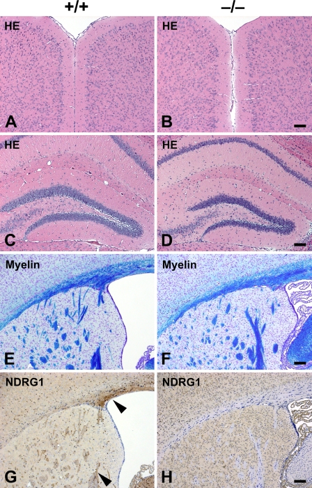
Figure 3.
Histological assessment of the brain of adult Ndrg1-deficient mice. Transverse sections of cerebral neocortex (A,B) and hippocampus (C,D) of adult wild-type (+/+; A,C) and adult Ndrg1-deficient (−/−; B,D) mice were compared. There were no significant differences in the structure of the brain regardless of genotype. Transverse sections of the forebrain at the level of the corpus callosum (E,F) and lateral ventricle (G,H) of adult wild-type (E,G) and adult Ndrg1-deficient (F,H) mice were compared. Normal myelination was observed in wild-type (E) and Ndrg1-deficient mice (F). NDRG1 was detected in the axon bundles of the corpus callosum and corpus striatum in wild-type mice (arrowheads in G). NDRG1 expression was not detected in the Ndrg1-deficient mice (H). HE, hematoxylin–eosin staining; myelin, luxol–fast blue staining; NDRG1, anti-NDRG1 immunostaining. Bar = 100 μm.

