Figure 1.
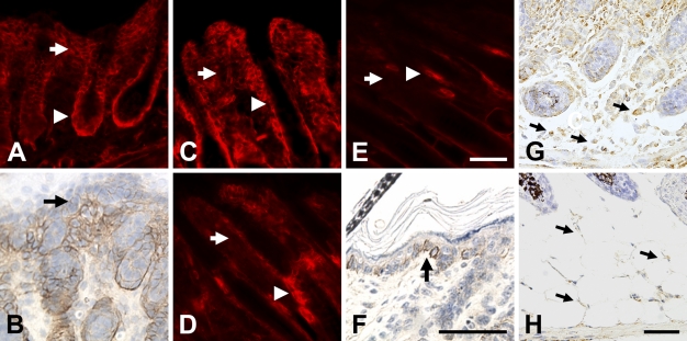
NG2 expression during early postnatal skin development. On postnatal day 1 (A), NG2 expression is strong in both the dermis (arrow) and outer root sheath of the hair follicle (arrowhead). Superficial to the dermis, immunoperoxidase staining reveals weaker expression of NG2 in the basal layer of keratinocytes (arrow in B). At day 3 (C), NG2 immunoreactivity remains strong in the hair follicle outer root sheath (arrowhead) but begins to diminish in the dermis (arrow). By days 7 (D) and 10 (E), NG2 has virtually disappeared from the dermis (arrow) and is mainly seen in the hair follicle bulge region (arrowheads). Immunoperoxidase labeling (F) reveals NG2 expression by basal keratinocytes at day 10 (arrow). In situ hybridization was used at postnatal days 1 (G) and 7 (H) to detect NG2 transcripts in adipocytes located in the subcutis (arrows); i.e., beneath the dermis and hair follicles. Bar = 50 μm.

