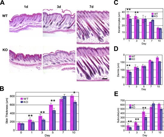
Figure 3.
Skin development in the NG2 null mouse. Images of hematoxylin- and eosin-stained skin sections (transverse) were prepared from wild-type (WT) and NG2 null mice during the first 10 days postnatally (A) and used to measure the overall skin thickness (B) and the thickness of the epidermis (C), dermis (D), and subcutis (E). At least six mice were used at each time point to establish the average values shown in the panels. Results were analyzed using the two-tailed Student's t-test. Statistically significant differences between WT and NG2 null samples are designated as follows: **p<0.01; *p<0.05. Bar = 200 μm.

