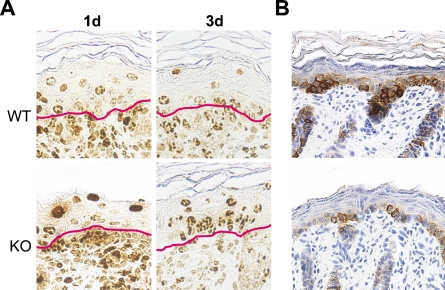
Figure 4.
Decreased keratinocyte production in the NG2 null mouse. (A) Following BrdU administration at 17 days of gestation, BrdU-labeled cells in the skin were visualized at days 1 and 3 by staining with anti-BrdU antibody. Red lines in each panel mark the dermis/epidermis boundary. (B) Immunoperoxidase labeling for CK-5 illustrates the deficiency in basal keratinocytes in NG2 null skin.

