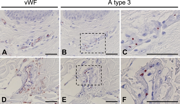
Figure 2.
Immunohistochemical staining of the skin of a 1-day-old wound (Group II). Vascular endothelial cells in the dermis (A) and subcutaneous region (D) of 1-day-old wound specimens were identified by immunostaining with anti-human von Willebrand factor (vWF) antibody. A type 3 antigens were strongly detected in vascular endothelial cells in the adjacent sections (B,E). Areas indicated by boxes in B and E are depicted at higher magnification in C and F, respectively. Bar = 50 μm.

