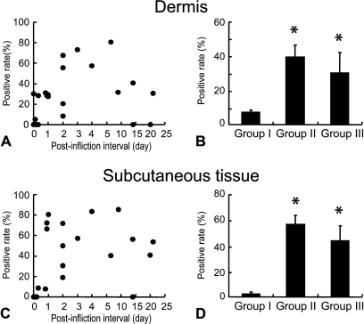
Figure 3.
Ratio of A type 3–positive vascular endothelial cells in the dermis (A) and subcutaneous tissue (C) in relation to postinfliction intervals. Vascular endothelial cells were identified by immunostaining of adjacent sections with anti-human vWF antibody as shown in Figure 2. Mean value and SE of A type 3–positive vascular endothelial cells in the dermis (B) and subcutaneous tissues (D) in each wound group. *Significant difference from Group I was observed statistically (p<0.05).

