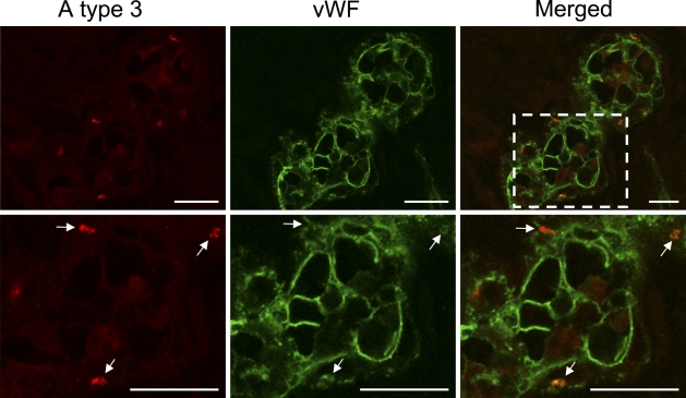
Figure 5.
Double-color immunofluorescence analysis of wounded skin tissues using a combination of AR-1 and antibody to vWF. A type 3 antigens (red) located in cells stained with anti-vWF (green) in 4-day-old wounded skin. Area indicated by box in merged panel is depicted at higher magnification in bottom panels. All AR-1–reactive spots were found in the regions reactive to anti-human vWF antibody. As shown in bottom panels, the discrete spots (white arrows) reactive to AR1 were completely merged to anti-human vWF-reactive spots. Bar = 20 μm.

