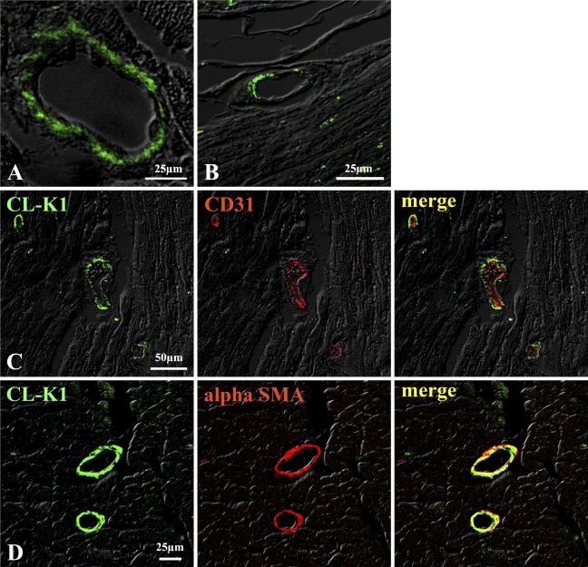
Figure 4.
IHC of vascular cells in heart (A) and small intestine (B). CL-K1 expression was detected in vascular portion in heart (A) and small intestine (B). Double immunofluorescence staining (C,D) demonstrates that CL-K1 was colocalized in vascular smooth muscle cells but not in endothelial cells.

