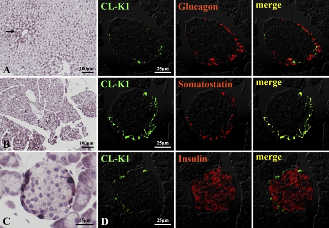
Figure 6.
IHC localization of CL-K1 in liver and pancreas. In liver (A), CL-K1 was expressed in hepatocytes. A relatively high expression of CL-K1 was seen in hepatocytes around the central vein (black arrow). In pancreas (B), CL-K1 was expressed not only in acinar cells but also in islet cells (C). Double immunofluorescence staining (D) demonstrates that CL-K1 was colocalized in somatostatin-containing D cells but not in glucagon-containing α-cells or insulin-containing β-cells.

