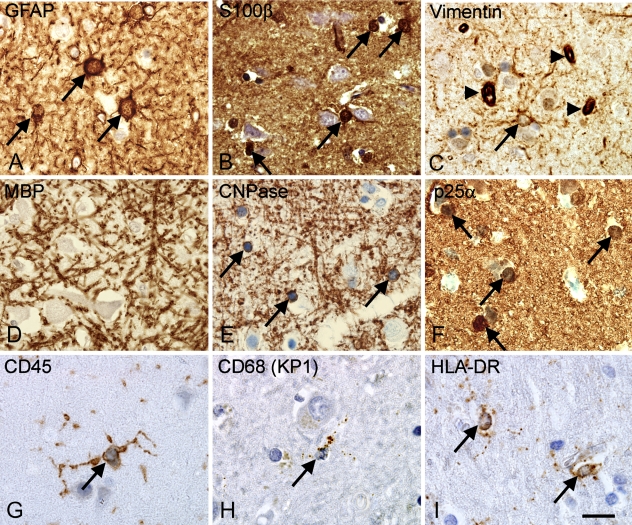
Figure 3.
Visualization of astroglial, oligodendroglial, and microglial cell bodies in the parietal neocortex from an adult donor. (A–C) Staining for astroglial markers showing glial fibrillary acidic protein (GFAP)+ astroglia (A), S100β+ glia (B), and vimentin+ astrocytes and cells of the capillary walls (arrowheads) (C). (D–F) Markers of oligodendroglia and myelin showing myelin basic protein (MBP)+ myelin fibers but no labeled cell bodies (D), 2′3′-cyclic nucleotide 3′-phosphodiesterase (CNPase)+ myelin fibers and round cell bodies (arrows) (E), and round cell bodies labeled by P25 α-antigen/tubulin polymerization promoting protein (p25α) (F). (G–I) Staining for microglia showing CD45+ ramified microglia (G), CD68+ (KP1) microglia showing a more punctuate labeling of their cell processes (H), and HLA-DR labeling the cellular processes (I). Paraffin sections from tissue array A were stained using the Dako ChemMate LSAB system, HIER method, and antibody dilutions listed in Table 5. Micrographs were acquired in neocortical layer VIa in samples from Donor 6 (Table 1). Labeled cell bodies are indicated by arrows. Bar = 20 μm.

