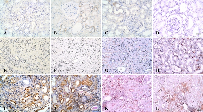
Figure 4.
Microphotographs of immunostaining for CD40, CD40L, TNF-α, and IL-1β in controls and patients with LN. (A–D) Normal controls. Weak immunoreactivity with CD40 (A), CD40L (B), and TNF-α (C) was detected in renal tubules of normal kidneys, whereas IL-1β expression was absent in the normal kidney tissue (D). (E–H) MCD. Expression of CD40 (E) and CD40L (F) was barely observed in the tubulointerstitium of patients with MCD, whereas TNF-α expression (G) was localized to the apical surface of proximal tubules, and expression of IL-1β (H) was present in the peritubular capillaries of a patient with MCD. (I–L) LN. Pronounced overexpression of CD40 (I), CD40L (J), TNF-α (K), and IL-1β (L) was observed in damaged tubules and interstitial infiltrates in patients with class IV-G LN. Bar (A–D; E–H,K,L; I,J) = 50 μm.

