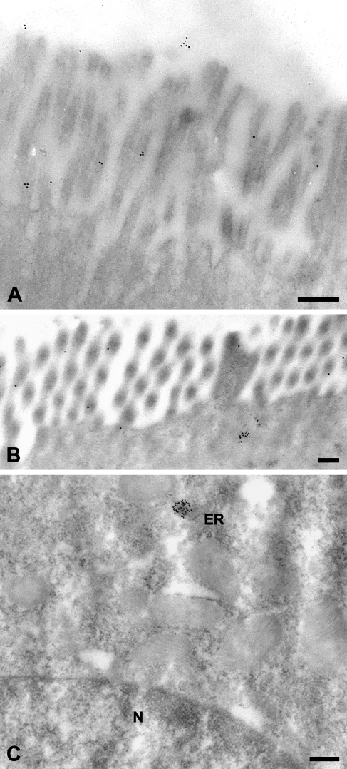
Figure 6.
Immune electron microscopic localization of the mCLCA6 protein. On the ultrastructural level, the mCLCA6 protein was localized to the brush border of enterocytes (A,B) in small clusters underneath the terminal actin web, in small clusters associated with the endoplasmic reticulum near the nucleus (C), and in small numbers in the lumen of the intestine (A). Tissue sections were incubated with αm6-N-1ap (diluted 1:12,000). Incubation with αm6-C-1bp (1:50) resulted in the same staining pattern except for the lumen, which was negative for the carboxy-terminal cleavage product. Incubation without primary antibody served as negative control. ER, endoplasmic reticulum; N, nucleus. Bar = 200 nm.

