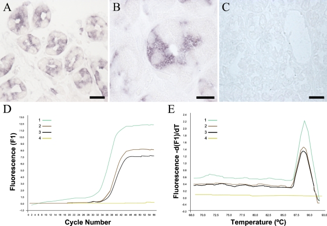
Figure 2.
CB1 mRNA is present in epithelial cells of the human gastric mucosa. (A,B) Hybridization with the antisense probe for CB1 mRNA followed by Tyramide signal amplification showed a strong signal in selective cells of the glandular epithelium, with morphological features coincident with the CB1-positive cells shown in Figure 1. Note the selective cytoplasmic distribution of the labeling, with nuclei lacking any staining. (C) Hybridization with the sense probe produced no significant staining. (D) Amplification curves of CB1 mRNA by RT-PCR in human cerebellum (Sample 1, positive control), human biopsies of gastric epithelium (Samples 2 and 3), and water (Sample 4, negative control). (E) Melting curves of the same samples as in D. Bars: A,C = 100 μm; B = 50 μm.

