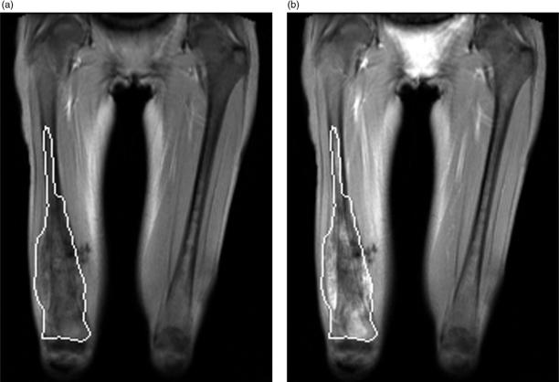
Figure 1.
Dynamic enhanced MRI examination of a 14-year-old girl with right femoral osteosarcoma. (a) Coronal T1 weighted image showing the region of interest (ROI) encompassing the entire tumor before contrast administration and (b) at the end of the 6-min scanning period. This tumor was a non-responder (<90% tumor necrosis).

