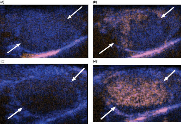
Figure 3.
Contrast enhanced ultrasound images of bevacizumab treated and control murine retroperitoneal neuroblastoma. (a) Before contrast administration and (b) at the peak of enhancement, this size matched control tumor (arrows) shows only minimal, peripheral perfusion (orange area). (c) Before contrast administration and at (d) peak enhancement, this tumor shows intense and homogenous perfusion 3 days after administration of one dose of bevacizumab.

