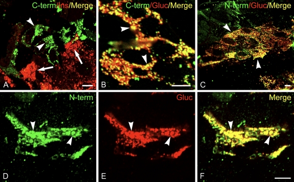
Figure 4.
CST-1 localizes to α cells in islets of Langerhans. Frozen thin sections of rat pancreas were stained with rabbit anti-CST-1 C-terminal (A,B) or N-terminal (C–F) antibodies and guinea pig anti-insulin (A) or glucagon (B–F) as well as Alexa 488-conjugated anti-rabbit IgG (green) and Alexa 594-conjugated anti-guinea pig IgG (red). CST-1 localized to glucagon granules in α cells (arrowheads) and did not stain insulin-containing β cells (arrows). CST-1 also localized to a few cells that did not label with glucagon, taken to be δ cells. Bar = 5 μm.

