Figure 4.
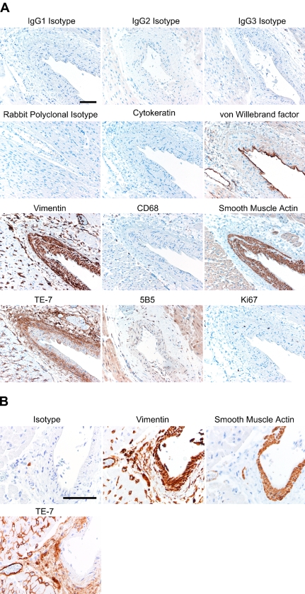
TE-7 recognizes adventitial fibroblasts surrounding blood vessels. (A) A portion of skeletal muscle from a healthy individual was formalin-fixed and paraffin-embedded. Serial sections were processed with a different primary antibody with its associated antigen retrieval method and secondary antibody. Results are shown for isotype controls (IgG1, IgG2, IgG3, and rabbit polyclonal). Staining with the AE1/AE3 antibody against epithelial cytokeratin was negative. Endothelial cells lining the blood vessels were recognized by an antibody against von Willebrand factor. All cells of mesenchymal origin, including fibroblasts and smooth muscle cells, were recognized by an anti-vimentin antibody. Cells of monocyte/macrophage lineage were scattered through the tissue and are identified by an anti-CD68 antibody. Smooth muscle cells of the tunica media labeled positively with an antibody to smooth muscle actin. Staining with the TE-7 antibody identified fibroblasts in the adventitia. The 5B5 antibody stained mostly surrounding muscle rather than fibroblasts. A small fraction of the cells are actively proliferating as determined by staining with an antibody that recognizes the proliferation marker Ki-67. (B) Heart muscle was formalin-fixed and paraffin-embedded. Serial sections were processed with antibodies as described in Materials and Methods. Staining with an isotype control IgG antibody resulted in little staining. An antibody to vimentin stained the tunica media, the adventitia, and cells within the heart muscle. An antibody to smooth muscle actin recognized the smooth muscle cells of the tunica media. Staining with the TE-7 antibody identified fibroblasts in the adventitia. TE-7 was largely negative on the tunica media smooth muscle cells. TE-7 also stained scattered cells and possibly a portion of the endomysium in the heart muscle. Bar = 10 μm.

