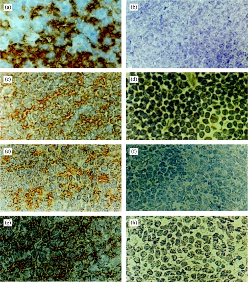
Figure 2.
Immunoperoxidase staining of frozen sections of normal human (a–d), mouse (e,f) and pig (g,h) thymus with either the mAb MR6 or the anti-gp200-MR6 specific phage antibody H4. This clone is used as a representative of the anti-gp200-MR6 antibodies isolated, which all gave similar staining results. The anti-gp200-MR6-specific phage antibody H4, labelled cortical epithelium from human (c), mouse (e) and pig (g) thymus with a similar pattern of staining as seen with mAb MR6 (a). The mAb MR6 did not stain mouse or pig thymus (data not shown). An irrelevant phage antibody anti-NIP (d,f,h) and the mAb OKT8 (b) were used as a negative control. Magnification ×800.

