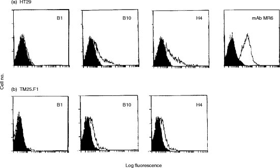
Figure 3.
Flow cytometric analysis of the human colon carcinoma cell line HT29 cells (a), the mouse thymic cortical epithelial cell line TM25.F1 (b) using either the mAb MR6 or anti-gp200-MR6-specific phage antibodies (B1, B10, H4). The shaded area represents background using the negative control antibody (OKT8 for MR6 or anti-NIP for phage antibodies). The mAb MR6 did not stain the TM25.F1 cell lines (data not shown).

