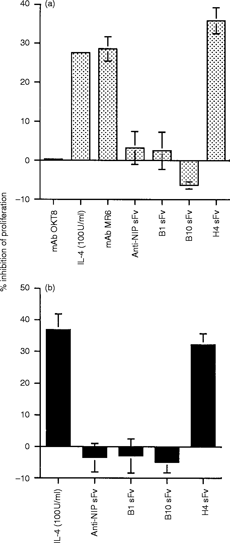
Figure 5.

The effect of mAb MR6, purified sFv and IL-4 on the proliferation of the human colon carcinoma cell line HT29 (a) and the mouse thymic cortical epithelial cell line TM25.F1 (b). HT29 cells were cultured either in medium alone or in medium supplemented with the experimental reagent for 48 hr and pulsed with [3H]thymidine for a further 8 hr and the amount of radioactivity (c.p.m.) incorporated was measured. The mAb MR6 and isotype-matched control OKT8 were used at 10 μg/ml. Recombinant human IL-4 was used at 100 U/ml. Purified sFv was used at 2 μg/ml. TM25.F1 cells were cultured either in medium alone or in medium supplemented with the experimental reagent for 24 hr and pulsed with [3H]thymidine for a further 8 hr and the amount of radioactivity (c.p.m.) incorporated was measured. Mouse IL-4 was used at 100 U/ml. Purified sFv was used at 2 μg/ml. The results are shown as percentage inhibition of proliferation and the values were estimated by comparing the difference in proliferation of cells cultured in medium supplemented, with the experiment reagent, against cells cultured in medium only. Columns and bars represent the mean±SD of triplicate determinants. In some columns the error bars are not visible due to the low value. For HT29 (a), the mAb MR6, IL-4 and H4 sFv and for TM25.F1 (b), IL-4 and H4 sFv significantly reduced cell proliferation; P < 0·01. The proliferation for HT29 and TM25.F1 cultured in medium only was 8·1×104 c.p.m. and 2·1×105 c.p.m., respectively.
