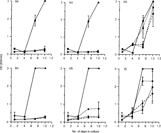
Figure 5.
FIV suppressor cell activity in WIV-vaccinated, protected cats. Mitogen-activated lymphoblasts were prepared from the blood (a and b), lymph nodes (c and d), and spleen (e and f) of two cats immunized with whole inactivated virus vaccine on three occasions, 3 weeks apart, 15 weeks following the final inoculation, and were cocultured with either FIVPET-infected (▪) or FIVGL-8-infected (•) MYA-1 cells at ratios of 4:1 (---), 2:1 (– – –), and 1:1 (· · ·). Control cultures contained FIV-infected MYA-1 cells alone (——). Replication of FIV was indicated by the detection of viral p24 in the culture supernatant by ELISA. The results shown represent the mean OD values at 650 nm measured for the two cats ± 2 SD, error bars are not discernible where the SD is small.

