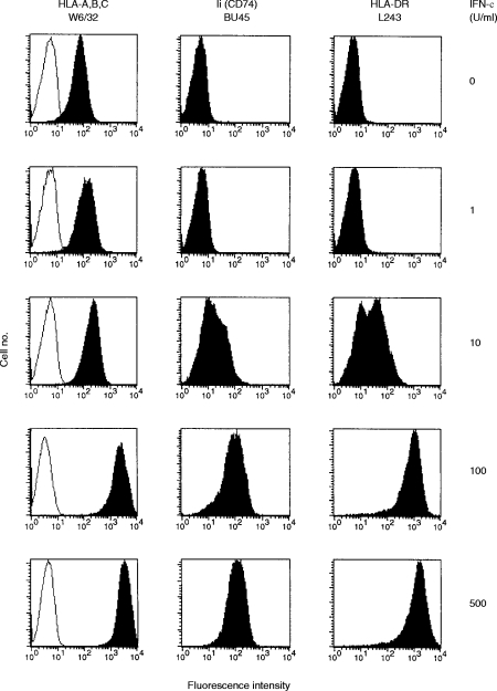
Figure 1.
Surface expression of major histocompatibility complex (MHC) class I molecules, Ii and human leucocyte antigen (HLA)-DR on interferon-γ (IFN-γ)-treated HT-29 cells. HT-29 cells cultured in the presence of the indicated concentrations of rIFN-γ (0–500 U/ml) for 72 hr were analysed by flow cytometry using the following mAbs: W6/32 (anti-HLA-ABC framework), BU45 (reacting with a C-terminal/extracellular determinant of all Ii isoforms) and L243 (reacting with mature HLA-DR molecules), represented by filled histograms, respectively. As a negative control, HT-29 cells were stained with the mAb VIC-Y1 (reacting with a cytoplasmic determinant of Ii, outlined histograms).

