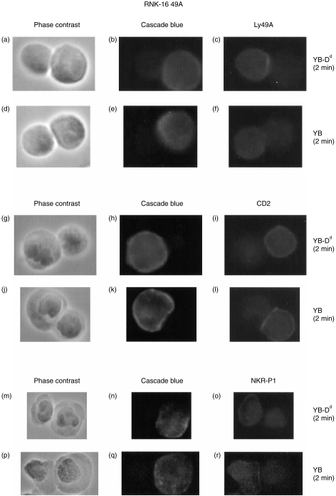
Figure 3.
Analysis of cell surface receptors on RNK-16 49A cells. RNK-16 49A cells were incubated for 2 min with YB-Dd (a–c), (g–i) and (m–o) or with YB (d–f), (j–l) and (p–r). The effector–target conjugates were then fixed in acetone (a–f) or in paraformaldehyde (g–r), and stained for Ly49A (c,f), CD2 (i,l) or NKR-P1 (o,r). (a, d, g, j, m and p) show phase contrast images of effector–target conjugates, and (b, e, h, k, n and q) show Cascade Blue© staining on target cells.

